المقالات
Osteosclerosis

Osteosclerosis is an abnormal change in the density of bone, not unlike a similar condition, condensing osteitis. However, there is often no obvious cause of osteosclerosis. Osteosclerosis lesions usually have a more dense appearance and show an irregular shape on x-ray examination. They also tend to be less reversible than condensing osteitis; as a result, they often are seen in areas of the jaws long after teeth have been extracted. They may result from a low-grade infection or persistent irritation to the tissue surrounding the tooth's root. The change in the bone associated with osteosclerosis may be part of the body's response to a low-grade infection, or even a reversible inflammation within the tooth's pulp.
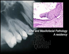
Osteosclerosis can also occur in response to deep decay in the tooth, large restorations, or injury to the tooth. The change in density can only be seen on a dental x-ray, where it appears as an enlarged white area. It usually occurs in the bone directly adjacent to the irritated tooth or teeth, or in the bone nearby where there has been a previous infection. The condition may involve any area of the upper or lower jaw bone, but is most common in the lower jaw, particularly in the area of the first molar. It can occur in anyone over three years of age, and typically causes no discomfort. It is most common in those over 20 years of age, and is not reversible. Fragments of the roots of primary or permanent teeth may be associated with osteosclerosis. The condition also often occurs at the same time as you experience heavy wear on your teeth, usually associated with tooth clenching and grinding.
No treatment is required, except for the removal of any obvious source of infection or irritation (such as abnormal chewing or biting habits).
* Treating the infection with antibiotics Once treatment is completed, the affected areas may remain unchanged for an indefinite period of time without causing you any problems. It's important that we keep an eye on the area by taking periodic x-rays.
