PATIENT 1InstructionLook at this patient's fundus.Salient featuresHistory· Family history (either autosomal recessive, which is most common, orautosomal dominant; the gene for both forms has been mapped to chromosome 16,Hum Mol Genet 1997; 6: 1823).- Upper gastrointestinal haemorrhage, myocardial intarctton, stroke andinter-mittent claudication, visual loss.· Hypertension (due to involvement of renal vasculature).ExaminationFundus shows angioid streaks, i.e. linear grey or dark red streaks with irregular edgeswhich lie beneath the retinal vessels (Figs 31 and 32; roughly 50% of patients withangioid streaks have pseudoxanthoma elasticum, whereas 85% of those withpseudoxanthoma have angioid streaks).Proceed as follows:· Look at the neck (Fig. 33), antecubital fossae, axillae (Fig. 34), groin andperi-umbilical region for loose 'chicken skin' appearance of skin.· Examine peripheral pulses (absent pulses due to peripheral arterial involvementbut acral ischaemia is uncommon due to development of collaterals).PATIENT 2InstructionLook at this patient.Salient featuresSmall yellow papules arranged in a linear or reticular pattern in plaques on the neck,axillae, cubital fossae, periumbilical region and groin.Proceed as follows:Tell the examiner that you would like to examine the fundus.DiagnosisThis patient has angioid streaks on fundoscopy and 'chicken-skin' appearance in theneck and axillae (lesions) due to pseudoxanthoma elasticum (aetiology).Read: N Engl J Med 1993; 333: 1240; N Engl J Med 1997; 337:828 (candidates areencouraged to refer to this picture for characteristic features of this disease).ADVANCED-LEVEL QUESTIONSIn which other conditions are angioid streaks seen?· Regularly seen in: Paget's disease, sickle cell disease.· Occasionally seen in: Ehlers-Danlos syndrome, hyperphosphataemia, leadpoisoning, trauma, pituitary disorders and intracranial disorders.What are angioid streaks caused by?They are caused by abnormal elastic tissue in the Bruch's membrane of the retina.Which fundal finding is virtually pathognomonic of pseudoxanthoma elasticum?'Leopard skin spotting' changes which consist of yellowish mottling of the posterior poletemporal to the macula. These may antedate angioid streaks.What is the triad of pseudoxanthoma elasticum, angioid streaks and vascularabnormalities known as?Groenblad-Strandberg syndrome.What are the cardiovascular manifestations of this condition?· Mitral valve prolapse.· Restrictive cardiomyopathy.· Renovascular hypertension.· Premature coronary artery disease resembling accelerated atherosclerosis dueto calcification of the internal elastic laminae of arteries. Arterial grafts should not beused for coronary artery bypass surgery in these patients because of possiblecalcification of the internal elastic laminae of the internal mammary artery.· Peripheral vascular disease.what are the gastrointestinal manifestations of this disease? Gastrointestinalhaemorrhage, particularly during the first decade. Bleeding compli-cations can beprevented by avoiding aspirin in patients with pseudoxanthoma elasticum.What are the causes of visual loss in pseudoxanthoma elasticum? Macular involvementby a streak; disciform scarring secondary to choroidal haemorrhage or traumaticmacular haemorrhage.What are the cutaneous histological features of pseudoxanthomaelasticum ? The histological diagnosis is made by doing 4 mm punch biopsy of scars orflexural skin of the neck or axillae in patients who have angioid streaks on fundoscopybut no visible skin lesions. The Verhoeff-van Gieson stain (for elastic tissue) revealscharacteristic fragmentation and clumping of elastic tissue in middle and deep dermis.The von Kossa stain (for calcium) shows staining of calcified elastic tissue in the middleand deep dermis. Calcified elastic tissue is the hallmark of pseudo-xanthoma elasticum.An arteriolar sclerosis develops in the media of muscular arteries and arterioles and asa result the lumen may become progressively and concentrically narrowed.K.W.L. Bruch (1819-1884), Professor of Anatomy at Giessen, Germany.E.E. Groenblad, Swedish ophthalmologist and J. Strandberg, Swedish dermatologist.'EL. Terry (1899-1946), Head of Ophthalmology at Harvard University, describedangioid streaks in Paget's disease.
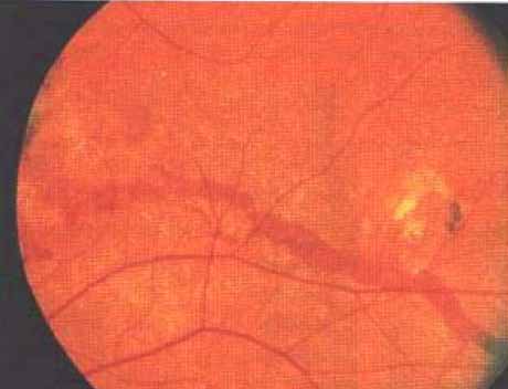
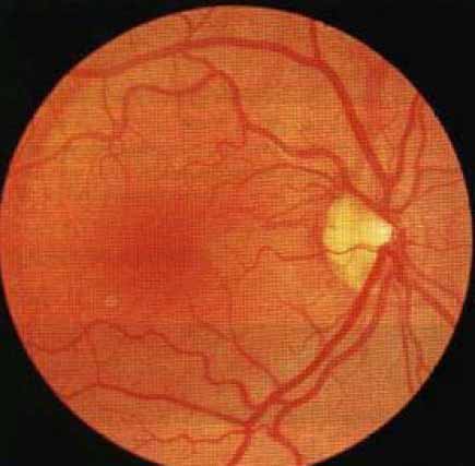 - Upper gastrointestinal haemorrhage, myocardial intarctton, stroke andinter-mittent claudication, visual loss.· Hypertension (due to involvement of renal vasculature).ExaminationFundus shows angioid streaks, i.e. linear grey or dark red streaks with irregular edgeswhich lie beneath the retinal vessels (Figs 31 and 32; roughly 50% of patients withangioid streaks have pseudoxanthoma elasticum, whereas 85% of those withpseudoxanthoma have angioid streaks).Proceed as follows:· Look at the neck (Fig. 33), antecubital fossae, axillae (Fig. 34), groin andperi-umbilical region for loose 'chicken skin' appearance of skin.
 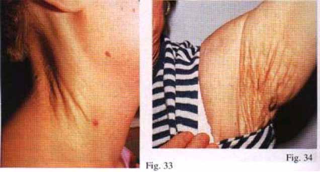 · Examine peripheral pulses (absent pulses due to peripheral arterial involvementbut acral ischaemia is uncommon due to development of collaterals).PATIENT 2InstructionLook at this patient.Salient featuresSmall yellow papules arranged in a linear or reticular pattern in plaques on the neck,axillae, cubital fossae, periumbilical region and groin.Proceed as follows:Tell the examiner that you would like to examine the fundus.DiagnosisThis patient has angioid streaks on fundoscopy and 'chicken-skin' appearance in theneck and axillae (lesions) due to pseudoxanthoma elasticum (aetiology).Read: N Engl J Med 1993; 333: 1240; N Engl J Med 1997; 337:828 (candidates areencouraged to refer to this picture for characteristic features of this disease).ADVANCED-LEVEL QUESTIONSIn which other conditions are angioid streaks seen?· Regularly seen in: Paget's disease, sickle cell disease.· Occasionally seen in: Ehlers-Danlos syndrome, hyperphosphataemia, leadpoisoning, trauma, pituitary disorders and intracranial disorders.What are angioid streaks caused by?They are caused by abnormal elastic tissue in the Bruch's membrane of the retina.Which fundal finding is virtually pathognomonic of pseudoxanthoma elasticum?'Leopard skin spotting' changes which consist of yellowish mottling of the posterior poletemporal to the macula. These may antedate angioid streaks.What is the triad of pseudoxanthoma elasticum, angioid streaks and vascularabnormalities known as?Groenblad-Strandberg syndrome.What are the cardiovascular manifestations of this condition?· Mitral valve prolapse.· Restrictive cardiomyopathy.· Renovascular hypertension.· Premature coronary artery disease resembling accelerated atherosclerosis dueto calcification of the internal elastic laminae of arteries. Arterial grafts should not beused for coronary artery bypass surgery in these patients because of possiblecalcification of the internal elastic laminae of the internal mammary artery.· Peripheral vascular disease.what are the gastrointestinal manifestations of this disease? Gastrointestinalhaemorrhage, particularly during the first decade. Bleeding compli-cations can beprevented by avoiding aspirin in patients with pseudoxanthoma elasticum.What are the causes of visual loss in pseudoxanthoma elasticum? Macular involvementby a streak; disciform scarring secondary to choroidal haemorrhage or traumaticmacular haemorrhage.What are the cutaneous histological features of pseudoxanthomaelasticum ? The histological diagnosis is made by doing 4 mm punch biopsy of scars orflexural skin of the neck or axillae in patients who have angioid streaks on fundoscopybut no visible skin lesions. The Verhoeff-van Gieson stain (for elastic tissue) revealscharacteristic fragmentation and clumping of elastic tissue in middle and deep dermis.The von Kossa stain (for calcium) shows staining of calcified elastic tissue in the middleand deep dermis. Calcified elastic tissue is the hallmark of pseudo-xanthoma elasticum.An arteriolar sclerosis develops in the media of muscular arteries and arterioles and asa result the lumen may become progressively and concentrically narrowed.K.W.L. Bruch (1819-1884), Professor of Anatomy at Giessen, Germany.E.E. Groenblad, Swedish ophthalmologist and J. Strandberg, Swedish dermatologist.'EL. Terry (1899-1946), Head of Ophthalmology at Harvard University, describedangioid streaks in Paget's disease.