المقالات
Pulp Stones (Denticles)

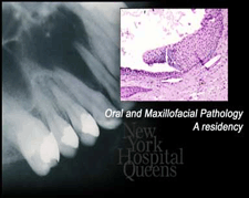
Pulp stones are hard, bone-like structures that form within the pulp of a tooth, either within the crown or within the root's canal. They are usually detected on x-ray examination, unless they are too small or are not dense enough to show up on an x-ray. In this case, the pulp stones are detected when we examine your tooth under a microscope after the tooth has been extracted (removed).
Pulp stones may exist as solitary or multiple bony formations within the pulp tissue, or they may be attached to the walls of the pulp chamber. Most appear to be attached when the extracted tooth is examined under a microscope. Pulp stones vary in size considerably, and in some cases, they appear to fill the pulp chamber or a portion of the root canals. Occasionally, they have been found in teeth that have not yet grown in.
Pulp stones are extremely common, occurring in as many as 90 percent of people between the ages of 50 and 70. Their incidence seems to increase with age. It is not known precisely what causes pulp stones, but they are not painful and typically present no symptoms or problems, other than occasionally getting in the way during root canal therapy.
Because most pulp stones are attached to the inner dentin wall, the irpresence can make root canal therapy very difficult to impossible. If
this is the case, and you are in need of root canal therapy to save a tooth, we will need to surgically remove the pulp stones.
